Following Cellular Lineage
Health & BehaviorA group of researchers based at UC San Diego and Rady Children's Institute have advanced the understanding of how the cerebral cortex develops by tracing the lineage of certain brain cells.

UC San Diego researchers and scientists are prominent in four projects selected by the Department of Defense to develop cutting-edge technologies.
A group of researchers based at UC San Diego and Rady Children's Institute have advanced the understanding of how the cerebral cortex develops by tracing the lineage of certain brain cells.
Traumatic experiences in early childhood can cause metabolic changes that impact muscle function later in life, according to new research co-authored by UC San Diego professor Anthony Molina.
A photographic exhibition opening the evening of Wednesday, April 24 at the Qualcomm Institute captures caves long-considered by the Maya to be entrances to the underworld.
UC San Diego alumnus David Loo (BS ’91) has zigzagged continents to earn a world-class education and launch global companies, including ServiceNow. Loo was honored recently as the Department of Computer Science and Engineering's distinguished alumnus.
The Washington Post, April 15
National Geographic, April 11
NewScientist, April 10
San Diego Magazine, April 9
Architectural Digest, April 7
Popular Science, April 1
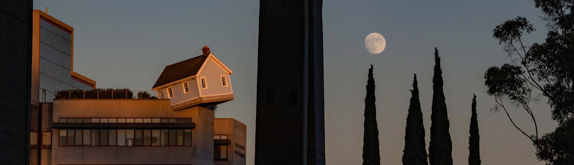
Take a deeper look into the remarkable stories of our changemakers and community-builders, our cutting-edge research, and the global influence of UC San Diego.
From exciting athletics programs and campus events, to our latest contributions to cutting-edge research—UC San Diego is a vibrant community of creators and innovators, making a positive impact both in San Diego and beyond.
Roger Hailstork recently rejoined UC San Diego as director of the UC San Diego Bookstore after a decade away. He is thrilled by the changes on campus—everything from new connections to the Divine Nine Plaza—and focused on making sure all students can get academic resources at an accessible price.
Satomi Rash-Zeigler will set the vision for the center and lead its strategic growth by fostering collaborations between faculty, students, workers and community organizations.
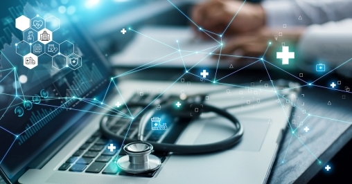
The University of California San Diego’s Steven M. Strauss and Lise N. Wilson Center for Cardiomyopathy will host the inaugural Cardiomyopathy Precision Medicine Symposium in La Jolla, Calif., on May 3, 2024.
“Prioritizing meaningful, deep connections with staff and students has been so impactful, and it’s wonderful getting to know people, learning what drives them and why they do what they do on such a diverse campus.”
“Together, we can build a world where we don’t have to question whether our basic human rights will be met. It takes hard work as a collective to try to battle systemic issues that are larger than us.”
“Connections with other students filled with big ideas, faculty who challenge us to aim higher and fellow alumni who share our desire to make change in the world have, for me, always been at the heart of UC San Diego's identity.”
Have an event to add to the UC San Diego Event Calendar? If you are a UC San Diego affiliate you can submit an event
Keep up with all the latest from UC San Diego. Subscribe to the newsletter today.