

A New View for Protein Turnover in the Brain
Researchers probe key processes potentially underlying a variety of neurological diseases
Published Date
By:
- Mario Aguilera
Share This:
Article Content
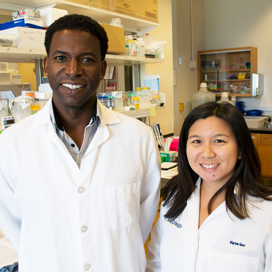
Gentry Patrick and Marisa Goo
Keeping the human brain in a healthy state requires a delicate balance between the generation of new cellular material and the destruction of old. Specialized structures known as lysosomes, found in nearly every cell in your body, help carry out this continuous turnover by digesting material that is too old or no longer useful.
Scientists have a strong interest in this degradation process since it must be tightly regulated to ensure healthy brain functioning for learning and memory. When lysosomes fail to do their job, brain-related disorders such as Parkinson’s and Alzheimer’s are possible.
Scientists at the University of California San Diego, led by graduate student Marisa Goo under the guidance of Professor Gentry Patrick, have provided the first evidence that lysosomes can travel to distant parts of neurons to branch-like areas known as dendrites. Surprisingly, they also found that lysosomes can be recruited to dendritic spines, specific areas where neurons communicate with each other. The researchers also revealed that direct activation of a single dendritic spine can directly recruit lysosomes to these specialized locations. The results are published in the Aug. 7 issue of the Journal of Cell Biology.
“Previously there was no reason to think that lysosomes could travel out to the ends of dendrites at synapses,” said Patrick a professor of neurobiology in UC San Diego’s Division of Biological Sciences. “We are showing neuronal activity is delivering them to the synapse and they are playing an integral and instructive role in remodeling and plasticity, which are so important for learning and memory.”
The researchers used genetically encoded fluorescent markers to label lysosomes and follow their movements, sometimes tens and even hundreds of microns away from the cell body. Confocal, two-photon, and electron microscopy were used to reveal that lysosomes move in dendrites and are present in spines, something previously unseen.
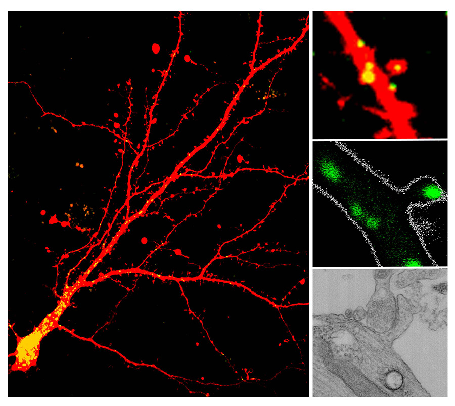
Lysosomes were identified in dendrites and dendritic spine using various techniques. Cultured neurons show lysosomes throughout neurons and in dendritic spines indicated in yellow (left and upper right); brain slices show a lysosome in the head of the spine highlighted in green (middle right); and transmission electron microscopy reveals a lysosome (black circle) near the base of a spine (bottom right).
“We’ve shown that lysosomes can be recruited to a single synapse… until now we had no idea that lysosomes could receive such instructive cues,” said Patrick, “For many neurodegenerative diseases, lysosome disfunction seems to play a role. So now we can look at the distribution and trafficking of lysosomes—which we now know are controlled by neurons—and ask: Is that altered in disease?”
Coauthors of the study include Laura Sancho, Natalia Slepak, Daniela Boassa, Thomas Deerinck, Mark Ellisman and Brenda Bloodgood.
The study was supported by a National Science Foundation graduate research fellowship (Goo), a Ford Foundation predoctoral fellowship (Goo), National Institutes of Health grant
NS060847 (Patrick), UC San Diego funds (Patrick), Searle Scholars program grant 14-SSP-184 (Bloodgood) and Pew Charitable Trusts grant 00028631 (Bloodgood). The National Center for Microscopy and Imaging Research at UC San Diego is supported by the National Institutes of Health grants P41GM103412 (Ellisman) and GM086197 (Stephen Adams/Ellisman/Boassa).
Share This:
You May Also Like


Stay in the Know
Keep up with all the latest from UC San Diego. Subscribe to the newsletter today.