

Nobel Laureate Roger Tsien Dies, Age 64
UC San Diego professor helped develop glowing proteins, illuminating life
Published Date
By:
- Scott LaFee
Share This:
Article Content
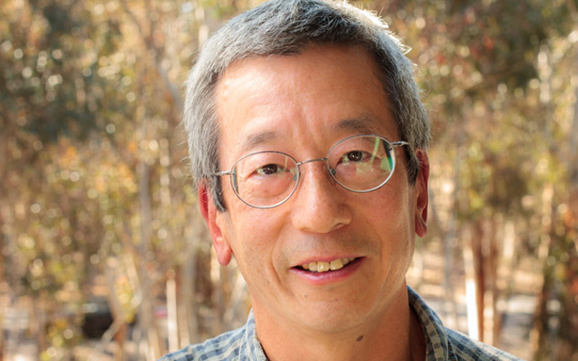
Roger Tsien, PhD, co-winner of the 2008 Nobel Prize in chemistry and professor of pharmacology, chemistry and biochemistry at University of California San Diego School of Medicine for 27 years, died August 24 in Eugene, Ore. He was 64.
Tsien’s work literally illuminated science. With Osamu Shimomura, PhD, an emeritus professor at the Marine Biological Laboratory in Woods Hole, Mass. and Martin Chalfie, PhD, a professor of biological sciences at Columbia University, Tsien helped scientists peer within living cells and organisms as never before, earning not just the 2008 Nobel Prize but scores of subsequent awards and accolades.
“Every honor was justly deserved, and always received with humility,” said Pradeep Khosla, chancellor of UC San Diego. “Roger was an extraordinary man: kind, generous, gracious, and always the consummate scientist pushing the limits of his work to expand the possibilities of science. He was a rare talent we cannot replace.”
Tsien, Shimomura and Chalfie collaborated to discover and develop green fluorescent protein (GFP), derived from the jellyfish Aequorea victoria, as a new and soon-indispensable research tool.
Shimomura identified the crucial jellyfish protein and revealed that it glowed bright green under ultraviolet light. Chalfie showed how it could be used as a biological marker. Combining his deep skills in chemistry and biology, Tsien found ways to make GFP glow more brightly and consistently; then he created a full palette of fluorescent proteins that scientists could use to track different cellular processes at the same time.
“I’ve always been attracted to colors,” Tsien told the San Diego Union-Tribune in 2008. “Color helps make the work more interesting and endurable. It helps when things aren’t going well. If I had been born color-blind, I probably never would have gone into this.”
GFPs have become a fundamental fixture in life sciences labs around the world, allowing researchers to look into cells or whole animals, to watch molecules interact in real-time and ask questions once thought impossible.
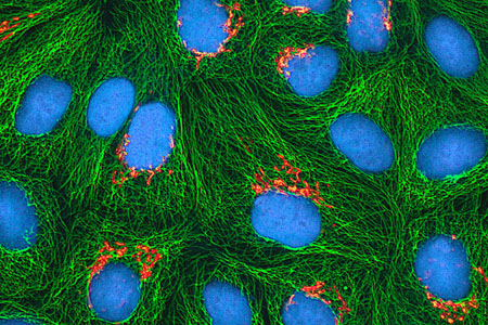
Cultured HeLa cancer cells depicted using fluorescent proteins to illustrate Golgi apparatus (orange) and microtubules (green), with DNA-carrying nuclei counterstained blue. Image courtesy of National Institutes of Health.
“Our work is often described as building and training molecular spies,” Tsien once said, “molecules that will enter a cell or organism and report back to us what the conditions are, what’s going on with the biochemistry, while the cell is still alive.”
Tsien was never content to rest upon his Nobel laurels. He wanted his research to be clinically relevant. Working with colleagues like Quyen T. Nguyen, MD, PhD, research collaborator and head and neck surgeon at UC San Diego Health, Tsien helped develop experimental injectable fluorescent peptides that cause hard-to-see peripheral nerves to glow, allowing surgeons to avoid them when removing damaged or cancerous tissues.
“The analogy I use is that when construction workers are excavating, they need a map showing where the existing underground cables are actually buried, not just old plans of questionable accuracy,” Tsien said. “Likewise when surgeons are taking out tumors, they need a live map showing where the nerves are actually located, not just a static diagram of where they usually lie in the average patient.”
As a distinguished Howard Hughes Medical Institute investigator, Tsien sought to better visualize cancer in other ways — or maybe treat it. He and colleagues have designed U-shaped peptides able to carry either imaging molecules or chemotherapy drugs to targeted cancer cells.
His lab created a new generation of fast-acting fluorescent dyes that optically highlight electrical activity in neuronal membranes, deciphering how brain cells function and interact. And using a modified plant protein, he and colleagues created a new type of genetic tag visible under an electron microscope (EM), allowing researchers to see life in unprecedented detail.
“The big advantage of EM is that it has always had much higher spatial resolution than light microscopy,” Tsien said. You can get up to a hundred-fold higher useful magnification from EM than from light microscopy.” The result is extraordinarily refined, three-dimensional images of microscopic objects at resolutions measuring in the tens of nanometers, tiny enough to meticulously render the internal anatomy of individual cells.
“Roger’s vision was vast and yet incredibly precise,” said David A. Brenner, MD, vice chancellor, UC San Diego Health Sciences and dean of UC San Diego School of Medicine. “He saw both the big picture, but also the incredible need to see and understand — in glorious color — all of the infinitesimal details that make it up, that make up life.”
“He was ahead of us all,” said Tsien’s wife, Wendy. “He was ever the adventurer, the pathfinder, the free and soaring spirit. Courage, determination, creativity and resourcefulness were hallmarks of his character. He accomplished much. He will not be forgotten.”
Biography in brief
Roger Yonchien Tsien was born February 1, 1952 in New York City, the third son of immigrant parents. He was a scientist from early childhood, sketching out chemistry experiments as an 8-year-old in a notebook now kept in the Nobel Museum in Stockholm, Sweden. His first Boy Scout merit badge was in chemistry.
In 1968 at the age of 16, he took first prize in the prestigious Westinghouse Science Talent Search for high school seniors. He attended Harvard College, graduating summa cum laude in chemistry and physics in 1972, then earned his doctorate in physiology in 1977 at the University of Cambridge in England.
Before coming to UC San Diego in 1989, he worked as a research assistant in Cambridge and then as a junior professor at UC Berkeley.
Tsien was a member of the Institute of Medicine, the American Academy of Arts and Sciences, the U.S. National Academy of Sciences and the Royal Society of London. Among his awards: the Gairdner Foundation International Award, the American Chemical Society Award for Creative Invention, the Heineken Prize for Biochemistry and Biophysics, the Max Delbruck Medal in Molecular Medicine, the Wolf Prize and the Keio Medical Science Prize.
Share This:

Stay in the Know
Keep up with all the latest from UC San Diego. Subscribe to the newsletter today.