
Acting Student and Bioengineering Alumna Awarded Soros Fellowship for New Americans
Awards & Accolades
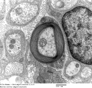
Nerve cell with protective layers of myelin.
In a paper published today in the journal Scientific Reports, a pair of researchers at the University of California, San Diego Skaggs School of Pharmacy and Pharmaceutical Sciences report that inhibiting the ability of immune cells to use fatty acids as fuel measurably slows disease progression in a mouse model of multiple sclerosis (MS).
MS is an autoimmune disease resulting from damage to the myelin sheath, a protective layer surrounding nerve cells. When the sheath is damaged, nerve impulses are slowed or halted, resulting in progressive physical and neurological disabilities. The cause of the damage is inflammation occurring when the body’s immune cells attack the central nervous system (CNS).
Marianne Manchester, PhD, professor of pharmacy and first author Leah P. Shriver, PhD, looked at how immune cells in the CNS oxidize fatty acids for energy when their preferred fuel source – glucose – is in short supply, which may occur in inflamed tissues. In a mouse model mimicking chronic MS, Manchester and Shriver discovered that by inhibiting a single enzyme that helps immune cells effectively exploit fatty acids, the cells eventually starved and died, preventing further inflammatory damage.
Currently, no approved drug or therapy for MS targets fatty acid metabolism. And the specificity of the target – inhibiting a single enzyme – suggests that adverse side effects associated with existing treatments, such as increased infection risk, is unlikely.
“We expect that because immune cells not in lesions in the CNS are able to use available glucose, they will function just fine during infection and that inhibition of this pathway would not produce general immune suppression,” Shriver said.
The enzyme-inhibitor used by Manchester and Shriver in their study is a drug already tested in humans with congestive heart failure, and was generally well-tolerated. The scientists are now using mass spectrometry to determine whether their results in the mouse model are translatable to humans. “We are interested in determining how this pathway is utilized in human tissue samples from MS patients,” Manchester said.
Funding for this study came from the National Institutes of Health and the National Institute of Neurological Disorders and Stroke.
Keep up with all the latest from UC San Diego. Subscribe to the newsletter today.

