
Acting Student and Bioengineering Alumna Awarded Soros Fellowship for New Americans
Awards & Accolades
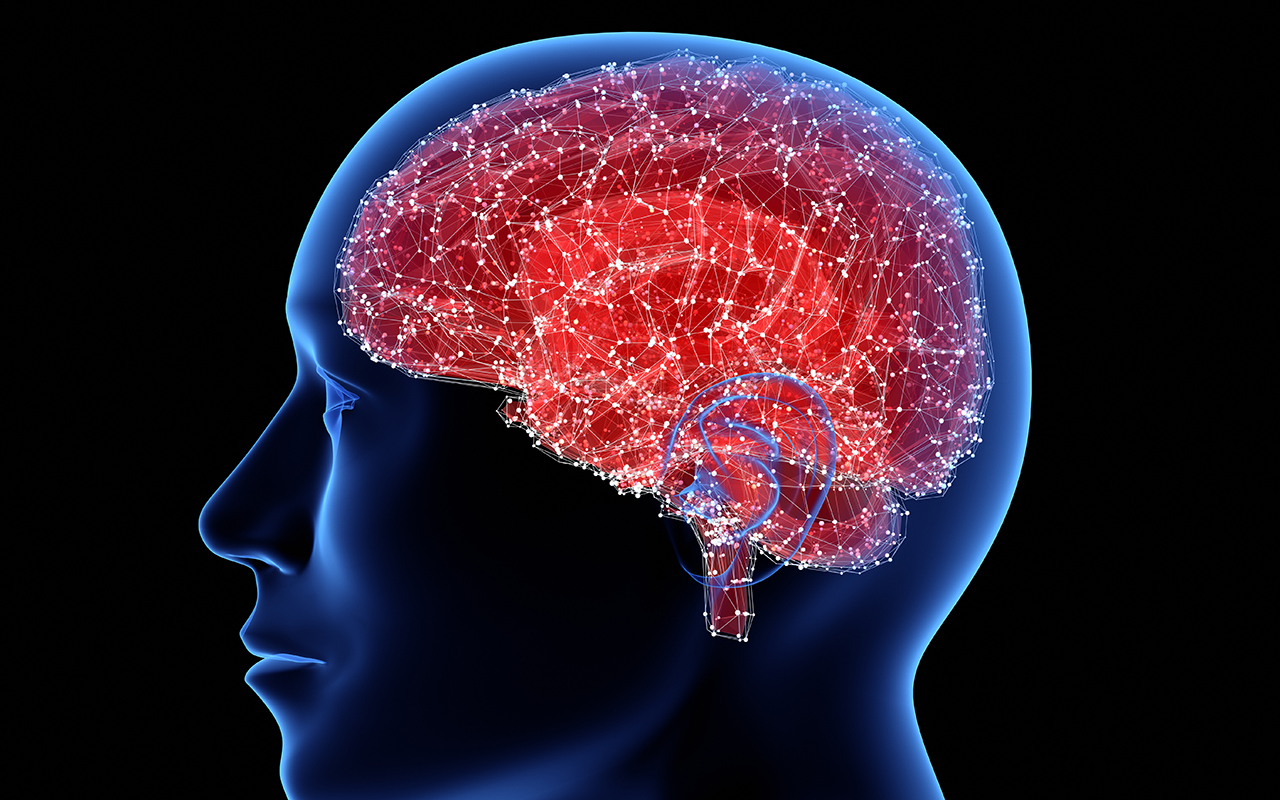
Researchers have developed new single-cell sequencing methods that could be used to map the cell origins of various brain disorders, including Alzheimer’s, Parkinson’s, schizophrenia and bipolar disorder.
By analyzing individual nuclei of cells from adult human brains, researchers at the University of California San Diego, Harvard Medical School and Sanford Burnham Prebys Medical Discovery Institute have identified 35 different subtypes of neurons and glial cells and discovered which of these subtypes are most susceptible to common risk factors for different brain diseases.
“There are multiple theories regarding the roots of various brain diseases. Our findings enable us to narrow down and rank which types of cells in the brain carry the most genetic risk for developing these diseases, which can help drug developers pick better targets in the future,” said Kun Zhang, a professor of bioengineering at the UC San Diego Jacobs School of Engineering and co-senior author of the study. Zhang is also a member of the Institute of Engineering in Medicine at UC San Diego.
This work builds off of a previous study published in Science, which Zhang also co-led, in which researchers identified 16 subtypes of neurons in the cerebral cortex. That study was the first large-scale mapping of gene activity in the human brain and provided a basis for understanding the diversity of individual brain cells.
“Our ultimate goal is to produce a complete cell atlas of the human brain,” Zhang said. “Here, we’ve created a fuller and more detailed map than what we’ve done in our previous work.”
In the new study, researchers developed a new generation of single-cell sequencing methods that enabled them to identify additional neuronal subtypes in the cerebral cortex as well as the cerebellum, and even further divide previously identified neuronal subtypes into different classes. The new methods also enabled researchers to identify different subtypes of glial cells, which wasn’t possible in the previous study due to the smaller size of glial cells.
“These data confirm and significantly expand our prior work, further highlighting the enormous transcriptional diversity among brain cell types, especially neurons,” commented co-senior author Jerold Chun, professor and senior vice president at Sanford Burnham Prebys Medical Discovery Institute. “This diversity, which continues to emerge from our single-cell analytical approach, will provide a foundation for better understanding the normal and diseased brain."
The advance was made possible by combining next-generation RNA sequencing with chromatin mapping—mapping of DNA and proteins in the nucleus that combine to form chromosomes—for more than 60,000 individual neurons and glial cells. The work was published Dec. 11 in Nature Biotechnology.
“While the analysis of RNA can tell us how cell types differ in their activity, the chromatin accessibility can reveal the regulatory mechanisms driving the distinctions between different cells”, noted Peter Kharchenko, an assistant professor of biomedical informatics at Harvard Medical School who co-led the study.
Using the information from RNA sequencing and chromatin mapping methods, researchers were able to map which cell types in the brain were affected by common risk alleles—snippets in DNA that occur more often in people with common genetic diseases. Researchers could then rank which subtypes of neurons or glial cells are more genetically susceptible to different brain diseases. For example, they found that two subtypes of glial cells, microglia and oligodendrocytes, were the first and second most at risk, respectively, for Alzheimer’s disease. They also identified microglia as most at risk for bipolar disorder, and a subtype of excitatory neurons as most at risk for schizophrenia.
“Now we can locate where the disease likely starts,” Zhang said. “However, we are only mapping the genetic risk. We don’t know the precise mechanism of how these specific cells actually trigger the disease.”
One caveat of this study, explained Zhang, is that it primarily analyzed data from adult brains (ages 20 to 50), so the findings do not represent younger or older populations. In order to better understand brain disorders that manifest early on, for example in infants, like autism spectrum disorder, the study would need to analyze cells from younger brains, he said.
The team also plans to expand their studies to map additional regions of the brain.
This work was supported in part by the NIH Common Fund Single Cell Analysis Program (1U01MH098977).
Paper title: “Integrative single-cell analysis of transcriptional and epigenetic states in the human adult brain.” Authors of the study are Blue B. Lake*, Song Chen*, Brandon C. Sos*, Thu E. Duong, Derek Gao and Kun Zhang of UC San Diego; Jean Fan* and Peter V. Kharchenko of Harvard Medical School; and Gwendolyn Kaeser, Yun C. Yung and Jerold Chun of Sanford Burnham Prebys Medical Discovery Institute.
*These authors contributed equally to this work.
Keep up with all the latest from UC San Diego. Subscribe to the newsletter today.

