

Borrowing from Astronomy to Rob the Twinkle from Brain Imagery
Neurophysicists make new recording of nerve signals by adapting tools of astronomers
Published Date
By:
- Cynthia Dillon
Share This:
Article Content
Image of a neuron in the output stage of a mouse cortex, filled with a dye that changes brightness according to the activity of the cell, together with a strip image of the angle of the whiskers. The activity, and thus brightness, of the neuron increases as the mouse activates muscles to protract the whiskers. Video courtesy of David Kleinfeld
For decades, astronomers have solved the problem of removing the “twinkle” from images of stars in the night sky. They use a process called “adaptive optics” (AO) to correct for changes in the density and moisture of air in the atmosphere that scatters the incoming star light.
Recently, UC San Diego Professor David Kleinfeld, along with postdoctoral fellow Rui Liu, realized how to adopt this same procedure to correct microscopic images for the scattering of light that occurs in brain tissue. The result of their adaptation was the first-ever recording of the subcellular neuronal inputs and outputs within the cortical mantle—the grey matter forming the cerebral cortex—in mice. Their study is currently published in Nature Methods.
According to Kleinfeld, this novel adaptation is likely to be a fundamental technical advance for the study of computing by nervous systems.
“Thus, as an example, we will be able to visualize how the sensation of touch is transformed into the movement of a limb,” said Kleinfeld, adding that the ability to characterize the input-output relation of circuits is an essential step in the study of any form of computing.
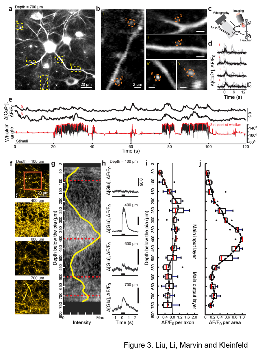
Imaging the microstructure of neuronal communication in the part of a mouse cortex that is sensitive to motion of the whiskers. (a,b) Imaging cell calcium, which reports activation of a neuron, in the output stage of the cortex. The expanded views show circled "spines,” which are the fundamental units of input to a cell, connected to "dendrites," the structures that integrate many inputs. (c) Cartoon that shows whisker tracking with a high-speed camera and air-puff stimulation to the whiskers. (d,e) The signaling to individual spines in panel b, measured through their Intracellular calcium (Ca2+) response, to air-puff stimulation of the one whisker. The whisker is held steady for the data of panel d. The mouse is freely whisking for the data of panel e; the concurrent angle (black) and set-point, or forward position (red) are shown. (f-j) Imaging of input signals to all layers in the cortex via the release of the neurotransmitter glutamate (glu) in response to air-puff stimulation of the vibrissa. Panel f shows the input fibers, called axons, for a cortical column, while panel g shows a projection of the fibers in the x-z plane along with the density (yellow) of fibers. Panel h shows the corresponding time-dependent signal of glutamate release, averaged over the region demarked by the red box in panel f. The glutamate response is shown as a population average of signal per axon in panel i. The same data, recalculated as signal per area, is shown in panel j. Figure courtesy of David Kleinfeld
The professor in the UC San Diego Department of Physics and Section of Neurobiology, who is a team leader in the National Institutes of Health’s Brain Research through Advancing Innovative Neurotechnologies® (BRAIN) Initiative, explained that the cortex is organized in terms of input, intermediate and output stages. The intermediate stage is on the surface of the cortex, making it most accessible, while the input stage is in the middle and the output stage on the bottom of the cortex—one millimeter deep in mice.
“Past work has focused almost exclusively on the top layer, or intermediate stage, although our UC Berkeley colleague Professor Na Ji used similar methods to probe the input stage,” noted Kleinfeld. “With the recent advance in AO microscopy at UC San Diego, we can now probe all stages—input, intermediate and output—to completely analyze how neuronal signals are transformed within the cortex.”
Other collaborators on the research involved Howard Hughes Medical Institute Investigator Jonathan Marvin and former UC San Diego undergraduate student Zengyi Li.
“It is a wondrous journey to be part of the neurophysics team at UC San Diego. Constructive discussions between Professor Kleinfeld, Zengyi and me are still quite vivid in my mind,” said Liu. “It was also a great opportunity to collaborate with Dr. Marvin, who provided critical help by synthesizing the indicators of neuronal signals. It was the constant guidance and support from my advisor and colleagues that made this project happen.”
The study was supported by a Major Research Instrumentation Award for instrument development from the National Sciences Foundation, as well as a Research Program Award from the National Institutes of Neurological Disease and Stroke. Design and fabrication of the microscope were supported by the National Science Foundation (MRI grant PHY153264) and physiological measurements were additionally supported by the National Institutes of Health (NINDS grant R35 NS097265).
Share This:
You May Also Like


Stay in the Know
Keep up with all the latest from UC San Diego. Subscribe to the newsletter today.