

Like Film Editors and Archaeologists, Biochemists Piece Together Genome History
Researchers uncover new evidence for the origin of RNA splicing within human genes
Published Date
By:
- Cynthia Dillon
Share This:
Article Content
Video shows the dynamics of the group II intron in action. Video courtesy of Daniel Haack
Old-school Hollywood editors cut out unwanted frames of film and patched in desired frames to make a movie. The human body does something similar—trillions of times per second—through a biochemical editing process called RNA splicing. Rather than cutting film, it edits the messenger RNA that is the blueprint for producing the many proteins found in cells.
In their exploration of the evolutionary origins and history of RNA splicing and the human genome, UC San Diego biochemists Navtej Toor and Daniel Haack combined two-dimensional (2D) images of individual molecules to reconstruct a three-dimensional (3D) picture of a portion of RNA—what the scientists call group II introns. In so doing, they discovered a large-scale molecular movement associated with RNA catalysis that provides evidence for the origin of RNA splicing and its role in the diversity of life on Earth. Their breakthrough research is outlined in a paper published in the current edition of Cell.
“We are trying to understand how the human genome has evolved starting from primitive ancestors. Every human gene has unwanted frames that are non-coding and must be removed before gene expression. This is the process of RNA splicing,” stated Toor, an associate professor in the Department of Chemistry and Biochemistry, adding that 15 percent of human diseases are the result of defects in this process.
Toor explained that his team works to understand the evolutionary origins of 70 percent of human DNA—a portion made up of two types of genetic elements, which are both thought to have evolved from group II introns. Specifically, spliceosomal introns, which make up about 25 percent of the human genome, are non-coding sequences that must be removed before gene expression. The other 45 percent is comprised of sequences derived from what are called retroelements. These are genetic elements that insert themselves into DNA and hop around the genome to replicate themselves via an RNA intermediate.
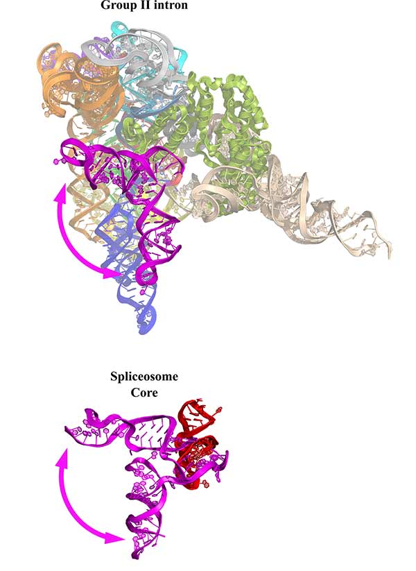
Figure 1 outlines the dynamics researchers see in the group II intron in comparison to the spliceosome. The figure highlights that both splicing machines undergo an identical 90-degree rotation of an important domain during splicing. Figure courtesy of Daniel Haack
“Studying group II introns gives us insight into the evolution of a large portion of the human genome,” noted Toor.
Working with the group II intron RNA nanomachine, Toor and Haack, a postdoctoral scholar at UC San Diego and first author of the paper, were able to isolate the group II intron complexes from a species of blue-green algae that lives at high temperature.
“Using a group II intron from a high-temperature organism facilitated structure determination due to the innate stability of the complex from this species,” said Haack. “The evolution of this type of RNA splicing likely led to the diversification of life on Earth.”
Haack further explained that he and Toor discovered that the group II intron and the spliceosome share a common dynamic mechanism of moving their catalytic components during RNA splicing.
“This is the strongest evidence to date that the spliceosome evolved from a bacterial group II intron,” he said.
Additionally, the findings reveal how group II introns are able to insert themselves into DNA through a process called retrotransposition. This copy-and-paste process has resulted in selfish retroelements proliferating in human DNA to comprise a large portion of the genome.
“Replication of these retroelements has played a large role in shaping the architecture of the modern human genome and has even been implicated in the speciation of primates,” noted Toor.
The researchers used cryo-electron microscopy (cryo-EM) to extract a molecular structure of the group II intron. They froze the RNA in a layer of thin ice and then shot electrons through this sample. According to the scientists, the electron microscope can magnify the image 39,000 times. The resulting 2D images of individual molecules were then put together to come up with a 3D view of the group II intron.
“This is like molecular archaeology,” described Haack. “Group II introns are living fossils that give us a glimpse into how complex life first evolved on Earth.”
This research was supported by the National Institutes of Health (grants 1R01GM123275, 1R01GM033050, NIH DP5 OD021396 and U54GM103368).
Share This:
You May Also Like


Stay in the Know
Keep up with all the latest from UC San Diego. Subscribe to the newsletter today.