

Magnets with ‘Magic Angle’ Offer New Way to Study Jelly States of Genes
Experimental innovation leads to useful information about structural changes in proteins
Published Date
By:
- Cynthia Dillon
Share This:
Article Content
Our cells all contain the same DNA and genes; but in different cells, different genes are switched on and off. This is why, for example, we have both blood cells and skin cells among others. This cellular switching is of interest to some scientists at UC San Diego who study molecular mechanisms of gene regulation in cells. Recent research by Assistant Professor of Chemistry and Biochemistry Galia Debelouchina ventured into this tiny atomic world of protein molecules within cells with a technological approach never before applied. The findings of her team’s study—recently published in “Angewandte Chemie, International Edition”—offer other researchers a new way to do their work.
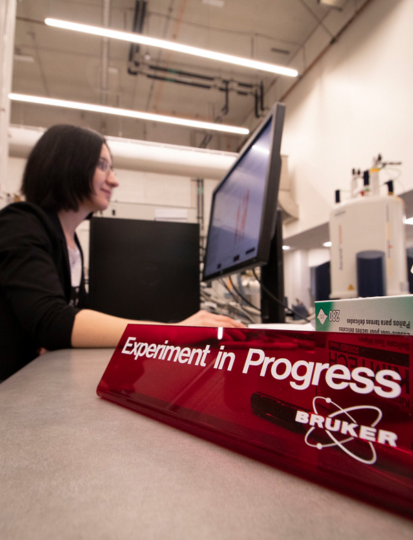
Professor Galia Debelouchina reviews data collected during the experiment. Photo by Michelle Fredricks, UC San Diego Physical Sciences
According to Debelouchina, biologists are working hard to understand how genes are regulated in the nucleus of a cell. Abnormal gene regulation can result in cells with mistaken identities, which can lead to cancer and premature cell death.
“Understanding these mechanisms will allow researchers to correct those mistaken identities and design molecules that can prevent cancer and premature aging mechanisms from happening much earlier in the process,” explained Debelouchina.
The scientist, who develops chemical and spectroscopic tools for structural biology of complex biological systems, is interested in the molecular decisions that cells make to determine which genes to turn on and off. Assisted by Bryce Ackermann, a graduate student in her research group whom she credits with the study’s novel approach, Debelouchina focused on a protein known as Heterochromatin Protein 1 alpha (HP1α). According to the professor, it not only plays an important role in deciding which genes are inactivated, it also features “very cool” biophysical properties. For example, in water, HP1α can form liquid-liquid droplets, much like oil droplets can separate in water solutions. Also, after some time, these liquid droplets can turn into gels.
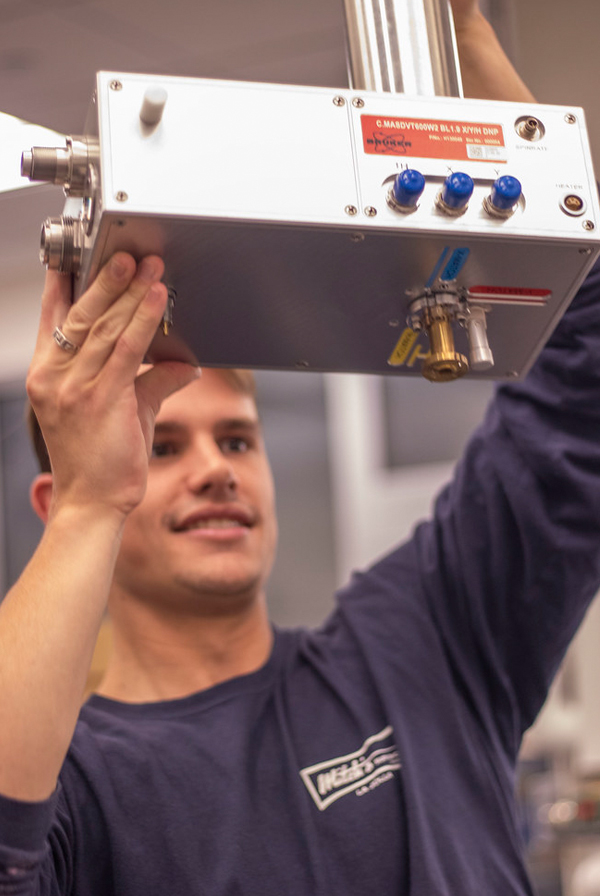
Bryce Ackermann displays the mechanism used to insert the rotor filled with sample into the magnet. Photo by Michelle Fredricks, UC San Diego Physical Sciences
“Many biologists believe that these properties are very important for its function in the cell,” said Debelouchina. “One hypothesis is that HP1α forms less mobile, jelly-like states around the genes to be inactivated, which prevents transcription factors from accessing those sites and turning the genes on. Our study shows two things: 1) we identify which parts of the protein are involved in the formation of the jelly-like state; 2) we show that other nuclear components such as chromatin, DNA wrapped around proteins, can modulate the jelly state, thus providing a way to regulate how and where it forms.”
Researchers are currently discovering many proteins that function in the cell through the formation of liquid-liquid droplets and gels. The sticky and unstructured traits of these substances, however, don’t allow for much useful information about the structural transformations of proteins using current structural biology techniques such as cryoEM, X-ray crystallography and solution nuclear magnetic resonance (NMR). But UC San Diego, according to Debelouchina, has excellent instrumentation resources for a different version of NMR called “magic angle spinning (MAS)” NMR. It can provide useful information for molecules in states similar to the ones that HP1α can adopt. So she and Ackermann added HP1α liquid droplets to tiny rotors that they inserted into large magnets—similar to those used for MRI experiments in hospitals, except they are designed to look into the atomic world of protein molecules instead of the components of human tissues.
“It was my first experiment with these instruments, so I had the thought that this approach of using liquid in a solid-state NMR experiment could work. The cool part was watching it form into gel over time,” said Ackermann. “It was rewarding to put theory into practice, see the stuff you read about and watch it work with your own hands.”
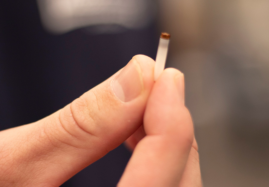
Graduate student Bryce Ackermann holds a rotor used in the experiment. Photo by Michelle Fredricks, UC San Diego Physical Sciences
Debelouchina admitted that she and Ackermann were surprised their technique worked and provided valuable information. She said they were also surprised by how much of an effect chromatin has on the gelation process of HP1α, adding that they will continue to explore the effect of other nuclear components in order to understand the delicate balance between liquid and jelly states in the nucleus.
“We thought that it would be interesting to try this technique, and it turned out to be tremendously useful,” said Debelouchina. “We were the first ones to ever try to get information in this way, and I suspect a lot of other researchers will now follow suit.”
The research was supported by a Technology Resource NIH grant (P41 EB002031) and by an NIH Molecular Biophysics Training Grant (T32 GM008326).Share This:
You May Also Like


Stay in the Know
Keep up with all the latest from UC San Diego. Subscribe to the newsletter today.