

Scientists Construct Google-Earth-like Atlas of the Human Brain
Published Date
By:
- Kim McDonald
Share This:
Article Content
Two neuroscientists have produced a new kind of atlas of the human brain that, they hope, can be eventually refined and improved to provide more detailed information about the organization and function of the human brain.
In an invited chapter published online this week in the Handbook of Clinical Neurology, Vol. 151, The Parietal Lobe, neuroscientists Ruey-Song Huang of UC San Diego and Martin Sereno of San Diego State University detail their surface-based human brain atlas.
The chapter and atlas are based on more than ten research papers the two researchers published in leading scientific journals, such as Nature Neuroscience, PNAS and the Journal of Neuroscience.
“Making a map is the first step in exploring a new world, whether it is an uncharted territory on the Earth or a new planet in outer space,” said Huang, a neuroscientist at UC San Diego’s Institute for Neural Computation. “For more than 25 years, noninvasive functional magnetic resonance imaging has allowed researchers to probe and map the human brain, one of the final frontiers, in unprecedented detail.”
He and Martin Sereno, the former chair of neuroimaging at University College London and a former professor at UC San Diego, said their atlas contains visual, somatosensory, multisensory, motor, action and motion maps that were pieced together from their own as well as other researchers’ functional magnetic resonance imaging, or fMRI, studies.
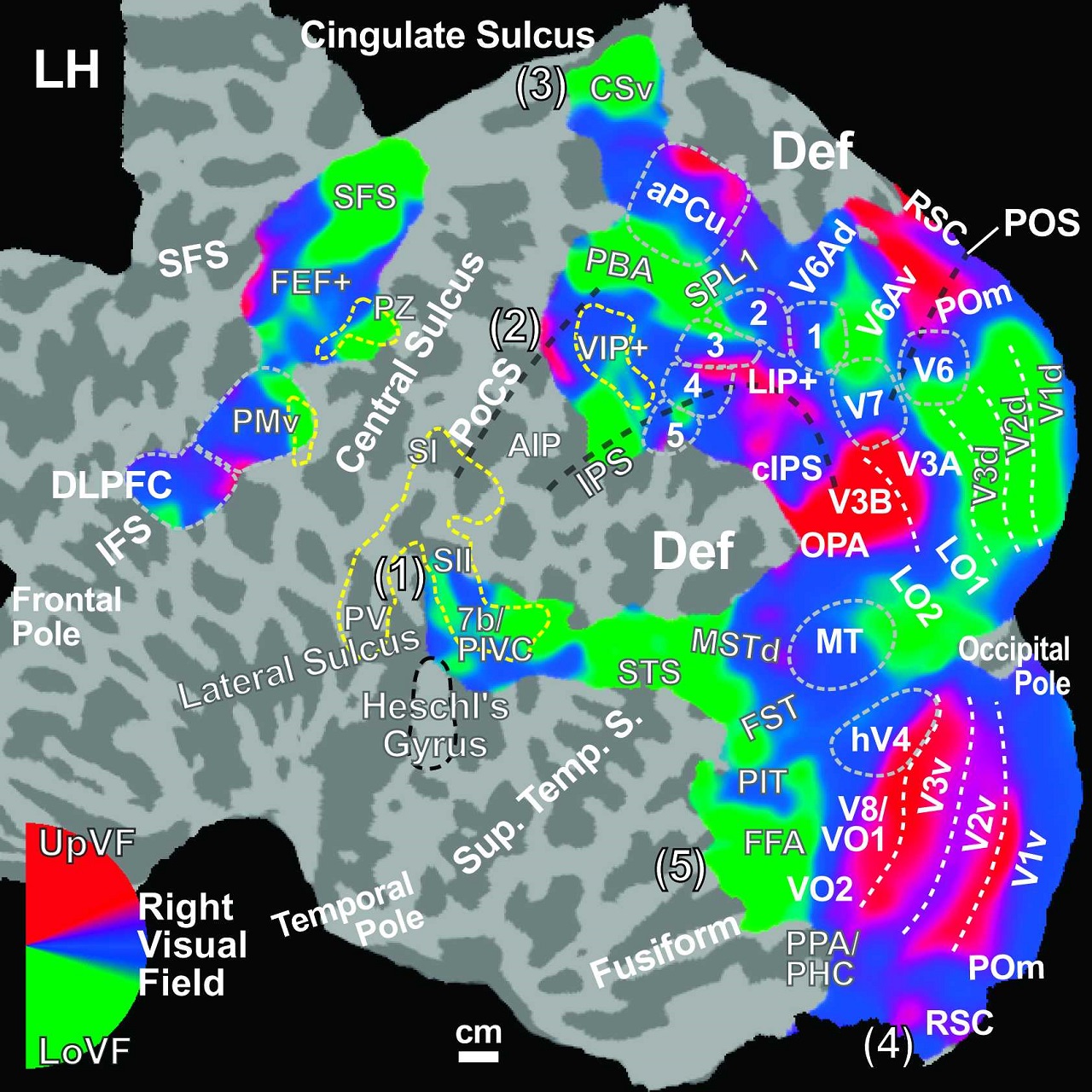
For neuroscientists, the two researchers said their new brain atlas reveals more than 40 “retinotopic areas” distributed across five visual streams — a more comprehensive picture of the human visual system than the initial division into a dorsal “where” pathway and a ventral “what” pathway. Full-body tactile stimulation and wide-field visual stimulation, they said, revealed a new multisensory homunculus located at the border between somatosensory and visual maps.
The scientists said their multilayer maps rendered on the same cortical surface—a map overlay method typically used in geographic information systems like “Google Earth”—provide insight into understanding how visual, somatosensory and motor maps partially overlap each other to support sensorimotor functions such as eye movements, pointing, reaching, grasping, eating, ducking and walking in daily life.
They expect that the areal and functional definitions in this atlas will be refined and updated by future studies using high-resolution brain imaging and more sophisticated stimuli and tasks tailored to regions with different specificity.
The researchers said their ultimate goal is to construct an online surface-based atlas containing layered maps of multiple modalities that can be used as a guide map to understand the topological organization, functions, and disorders of the human brain.
This online atlas will be constructed for searching and browsing brain areas and functions, they said, include interactive multi-layer features similar to “Google Earth.” These include spherical surface coordinates, gyri/sulci, boundaries of areas (“GPS coordinates,” “geographical features” and “county lines”), and embedded links to publications and figures (“Google Scholar” and “Google Images”), all on the same interface.
The research effort was supported by the National Institutes of Health (R01 MH081990), a Royal Society Wolfson Research Merit Award and the Wellcome Trust.
Share This:
You May Also Like


Stay in the Know
Keep up with all the latest from UC San Diego. Subscribe to the newsletter today.